ID:GLSK_HUMAN DESCRIPTION: RecName: Full=Glutaminase kidney isoform, mitochondrial; Short=GLS; EC=3.5.1.2; AltName: Full=K-glutaminase; AltName: Full=L-glutamine amidohydrolase; Flags: Precursor; FUNCTION: Catalyzes the first reaction in the primary pathway for the renal catabolism of glutamine. Plays a role in maintaining acid-base homeostasis. Regulates the levels of the neurotransmitter glutamate in the brain. Isoform 2 lacks catalytic activity. CATALYTIC ACTIVITY: L-glutamine + H(2)O = L-glutamate + NH(3). ENZYME REGULATION: Isoform 1 and isoform 3 are activated by phosphate. Inhibited by BPTES. BPTES binds between subunits and favors dissociation of the tetramer into dimers. BIOPHYSICOCHEMICAL PROPERTIES: Kinetic parameters: KM=1.9 mM for glutamine (isoform 1); KM=1.4 mM for glutamine (isoform 3); SUBUNIT: Heterotetramer. Interacts with ATCAY; the interaction is direct and may control GLS localization, negatively regulating its activity. SUBCELLULAR LOCATION: Isoform 1: Cytoplasm, cytosol. SUBCELLULAR LOCATION: Isoform 3: Mitochondrion. TISSUE SPECIFICITY: Isoform 1 and isoform 3 are detected in brain cortex. Isoform 3 is highly expressed in astrocytoma, ganglioglioma and ependymoma. Isoform 1 is highly expressed in brain and kidney, but not detected in liver. Isoform 3 is highly expressed in heart and pancreas, detected at lower levels in placenta, lung, pancreas and kidney, but is not detected in liver. Isoform 2 is expressed in cardiac and skeletal muscle. SIMILARITY: Belongs to the glutaminase family. SIMILARITY: Contains 2 ANK repeats. CAUTION: Isoform 3 is predicted to be expressed at very low levels due to a premature stop codon in the mRNA, leading to nonsense- mediated mRNA decay (PubMed:14759258), but has been shown to be well expressed (PubMed:17940881 and PubMed:11015561) and the encoded protein is detected in mitochondria (PubMed:22228304). SEQUENCE CAUTION: Sequence=BAA74861.2; Type=Erroneous initiation; Note=Translation N-terminally shortened;
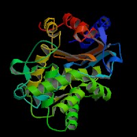
ModBase Predicted Comparative 3D Structure on O94925
Front
Top
Side
The pictures above may be empty if there is no ModBase structure for the protein. The ModBase structure frequently covers just a fragment of the protein. You may be asked to log onto ModBase the first time you click on the pictures. It is simplest after logging in to just click on the picture again to get to the specific info on that model.