ID:CEBPB_HUMAN DESCRIPTION: RecName: Full=CCAAT/enhancer-binding protein beta; Short=C/EBP beta; AltName: Full=Liver activator protein; AltName: Full=Nuclear factor NF-IL6; AltName: Full=Transcription factor 5; Short=TCF-5; FUNCTION: Important transcriptional activator in the regulation of genes involved in immune and inflammatory responses. Specifically binds to an IL-1 response element in the IL-6 gene. NF-IL6 also binds to regulatory regions of several acute-phase and cytokines genes. It probably plays a role in the regulation of acute-phase reaction, inflammation and hemopoiesis. The consensus recognition site is 5'-T[TG]NNGNAA[TG]-3'. Functions in brown adipose tissue (BAT) differentiation (By similarity). Regulates the transcriptional induction of peroxisome proliferator-activated receptor gamma (PPARG). SUBUNIT: Binds DNA as a dimer and can form stable heterodimers with C/EBP alpha, delta and gamma. Interacts with TRIM28 and PTGES2. Interacts with PRDM16. Interacts with CCDC85B. Forms a complex with THOC5 (By similarity). Interacts with ZNF638; this interaction increases transcriptional activation (By similarity). Interacts with CIDEA and CIDEC; these interactions increase transcriptional activation of a subset of CEBPB downstream target genes (By similarity). Interacts with DDIT3/CHOP. INTERACTION: P04637:TP53; NbExp=4; IntAct=EBI-969696, EBI-366083; SUBCELLULAR LOCATION: Nucleus. TISSUE SPECIFICITY: Expressed at low levels in the lung, kidney and spleen. INDUCTION: By ER stress. DOMAIN: the 9aaTAD motif is a transactivation domain present in a large number of yeast and animal transcription factors. PTM: Sumoylated by polymeric chains of SUMO2 or SUMO3. PTM: Phosphorylated at Thr-235 by RPS6KA1. SIMILARITY: Belongs to the bZIP family. C/EBP subfamily. SIMILARITY: Contains 1 bZIP (basic-leucine zipper) domain. WEB RESOURCE: Name=SeattleSNPs; URL="http://pga.gs.washington.edu/data/cebpb/";
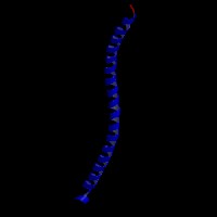
ModBase Predicted Comparative 3D Structure on P17676
Front
Top
Side
The pictures above may be empty if there is no ModBase structure for the protein. The ModBase structure frequently covers just a fragment of the protein. You may be asked to log onto ModBase the first time you click on the pictures. It is simplest after logging in to just click on the picture again to get to the specific info on that model.