ID:SP100_HUMAN DESCRIPTION: RecName: Full=Nuclear autoantigen Sp-100; AltName: Full=Lysp100b; AltName: Full=Nuclear dot-associated Sp100 protein; AltName: Full=Speckled 100 kDa; FUNCTION: May play a role in the control of gene expression. SUBUNIT: Homodimer. Splice variants heterodimerize. Interacts with members of the HP1 family of nonhistone chromosomal protein, such as CBX5 and CBX3 via the PxVxL motif. Interacts with Epstein-Barr virus EBNA-LP. Interacts with human cytomegalovirus/HHV-5 protein UL123. INTERACTION: P55854:SUMO3; NbExp=2; IntAct=EBI-751145, EBI-474067; SUBCELLULAR LOCATION: Nucleus, PML body. Note=Found in the nuclear body, also known as nuclear domain 10 (ND10), PML oncogenic domain (POD), nuclear dots (ND) and KR body. The nuclear body is a nucleoplasmic structure of punctate shape, which varies in size and number. Induction by interferon and may be cell cycle stages modulate the subnuclear localization of the isoforms. TISSUE SPECIFICITY: Widely expressed. Sp100-B is expressed only in spleen, tonsil, thymus, mature B-cell line and some T-cell line, but not in brain, liver, muscle or non-lymphoid cell lines. INDUCTION: By interferon. DOMAIN: The HSR domain is important for the nuclear body targeting as well as for the dimerization. DOMAIN: Contains one Pro-Xaa-Val-Xaa-Leu (PxVxL) motif, which is required for interaction with chromoshadow domains. This motif requires additional residues -7, -6, +4 and +5 of the central Val which contact the chromoshadow domain. PTM: Sumoylated. Sumoylation depends on a functional nuclear localization signal but is not necessary for nuclear import or nuclear body targeting. MISCELLANEOUS: The major isoform Sp100-A, has a calculated MW of 54 kDa, but exhibits aberrant electrophoretic mobilities, with an apparent MW OF 100 kDa. SIMILARITY: Contains 2 HMG box DNA-binding domains. SIMILARITY: Contains 1 HSR domain. SIMILARITY: Contains 1 SAND domain.
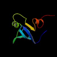
ModBase Predicted Comparative 3D Structure on P23497
Front
Top
Side
The pictures above may be empty if there is no ModBase structure for the protein. The ModBase structure frequently covers just a fragment of the protein. You may be asked to log onto ModBase the first time you click on the pictures. It is simplest after logging in to just click on the picture again to get to the specific info on that model.