Press "+" in the title bar above to open this section.
Comments and Description Text from UniProtKB
ID:DUT_HUMAN DESCRIPTION: RecName: Full=Deoxyuridine 5'-triphosphate nucleotidohydrolase, mitochondrial; Short=dUTPase; EC=3.6.1.23; AltName: Full=dUTP pyrophosphatase; Flags: Precursor; FUNCTION: This enzyme is involved in nucleotide metabolism: it produces dUMP, the immediate precursor of thymidine nucleotides and it decreases the intracellular concentration of dUTP so that uracil cannot be incorporated into DNA. CATALYTIC ACTIVITY: dUTP + H(2)O = dUMP + diphosphate. COFACTOR: Magnesium. ENZYME REGULATION: Phosphorylation is necessary for activity. BIOPHYSICOCHEMICAL PROPERTIES: Kinetic parameters: KM=2.5 uM for dUTP; Note=for both isoform 2 and isoform 3; PATHWAY: Pyrimidine metabolism; dUMP biosynthesis; dUMP from dCTP (dUTP route): step 2/2. SUBUNIT: Homotrimer. INTERACTION: Q6ZVK8:NUDT18; NbExp=3; IntAct=EBI-353224, EBI-740486; SUBCELLULAR LOCATION: Isoform 2: Nucleus. SUBCELLULAR LOCATION: Isoform 3: Mitochondrion. TISSUE SPECIFICITY: Found in a variety of tissues. Isoform 3 expression is constitutive, while isoform 2 expression correlates with the onset of DNA replication (at protein level). Isoform 2 degradation coincides with the cessation of nuclear DNA replication (at protein level). PTM: Nuclear isoform 2 is phosphorylated in vivo on Ser-11, a reaction that can be catalyzed in vitro by CDC2. Phosphorylation in mature T-cells occurs in a cell cycle-dependent manner. Isoform 3 is not phosphorylated. PTM: The initiator methionine is cleaved in isoform 2. MISCELLANEOUS: Each trimer binds three substrate molecules. The ligands are bound between subunits, and for each substrate molecule, residues from adjacent subunits contribute to the binding interactions. SIMILARITY: Belongs to the dUTPase family. SEQUENCE CAUTION: Sequence=AAB71393.1; Type=Frameshift; Positions=29, 47; Sequence=AAB93866.1; Type=Frameshift; Positions=29, 47; Sequence=AAB94642.1; Type=Frameshift; Positions=29, 47; WEB RESOURCE: Name=NIEHS-SNPs; URL="http://egp.gs.washington.edu/data/dut/";
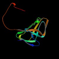
ModBase Predicted Comparative 3D Structure on P33316
Front
Top
Side
The pictures above may be empty if there is no ModBase structure for the protein. The ModBase structure frequently covers just a fragment of the protein. You may be asked to log onto ModBase the first time you click on the pictures. It is simplest after logging in to just click on the picture again to get to the specific info on that model.